
Mass spectrometry: SIMS
Secondary Ion Mass Spectrometry (SIMS) is a highly sensitive surface analysis technique that identifies the elemental, isotopic and molecular composition of a material. A focused primary ion beam releases secondary ions from the sample surface, which are then analysed. SIMS can be performed in static, dynamic or imaging modes, depending on the application.
Introduction
Secondary Ion Mass Spectrometry (SIMS) analyses the outermost surface of a material by directing a focused ion beam onto the sample. This releases secondary ions, which are measured to identify the elemental, isotopic and molecular composition of the surface. SIMS is widely used to analyse thin films, coatings and surface contamination, and can detect almost all elements, including hydrogen, at very low concentrations.
Secondary Ion Mass Spectrometry (SIMS) uses a focused primary ion beam, such as Cs⁺, O₂⁺, Ga⁺ or cluster sources like Bi or C₆₀, to sputter material from a sample surface. A fraction of the sputtered species are emitted as secondary ions and extracted into a mass spectrometer, where their mass-to-charge ratio is measured using systems such as quadrupole, magnetic sector or time-of-flight analysers.
Static SIMS operates at low primary ion doses to retain molecular information from the outermost surface layer. Dynamic SIMS uses higher ion doses for depth profiling through bulk material. Imaging SIMS scans the beam across the surface to generate spatially resolved elemental and molecular maps, with nanometre-scale lateral resolution available on NanoSIMS and high-resolution ToF-SIMS instruments.
In Secondary Ion Mass Spectrometry (SIMS), sensitivity is governed by sputtering yield, ionisation probability and matrix effects, all of which depend on the primary ion species, energy and sample composition. Cluster and polyatomic ion beams such as C₆₀, large argon clusters or water clusters reduce subsurface damage and increase molecular ion yield, enabling more effective analysis of organic and biological materials.
In time-of-flight SIMS, mass resolution is influenced by the initial kinetic energy spread of the secondary ions and the timing of the extraction field. Reflectron optics and delayed extraction are used to correct for energy dispersion and improve peak resolution. Detector response, including dead time and dynamic range, also places practical limits on quantification at high count rates.
Secondary Ion Mass Spectrometry (SIMS) provides direct molecular and fragment ion information from a surface without the need for derivatisation, labelling or extensive sample preparation. It is widely used to study surface chemistry, including self-assembled monolayers, thin films, coatings and polymer or material interfaces.
Ionisation efficiency in SIMS is strongly matrix-dependent, so quantification typically relies on matched standards or relative sensitivity factors. The use of cluster primary ion sources has significantly improved chemical information, enabling detection of larger organic molecules with reduced fragmentation compared with monatomic ion bombardment.
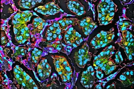
Secondary Ion Mass Spectrometry (SIMS) imaging maps the chemical composition of cells, tissue sections and microbial samples without the need for staining or labelling. It can localise lipids, drugs, metabolites and isotopically labelled tracers directly within sample structures, with spatial resolution down to tens of nanometres using NanoSIMS.
This makes SIMS a strong complement to fluorescence and electron microscopy in studies of single-cell metabolism, drug distribution in tissue, and microbial or biofilm chemistry, where label-free, spatially resolved molecular information is required.
Key features
Areas of use
SIMS is used any time someone needs to know exactly what is sitting on, or just below, the surface of a material:
- Checking a coating or thin film for contamination before it goes into a product
- Working out why a component failed by analysing its surface chemistry
- Mapping where a drug or chemical has reached inside a tissue sample
- Confirming the thickness and composition of layers in an electronic device
- Detecting trace levels of dopants or impurities in semiconductor materials
- Semiconductor metrology: Dopant and impurity depth profiling, junction analysis and contamination screening in device fabrication.
- Materials science: Thin-film composition, interface chemistry and coating uniformity across multilayer structures.
- Failure analysis: Root-cause investigation of surface-related defects, delamination and corrosion-driving contaminants.
- Battery research: Characterisation of electrode-electrolyte interfaces and degradation products in lithium-ion and solid-state cells.
- Biological imaging: Label-free molecular mapping of lipids, drugs and metabolites in cells and tissue sections.
- Primary ion source development: Characterisation of cluster and polyatomic ion sources for improved sputtering yield and reduced damage.
- Ion optics and mass analyser design: Optimisation of extraction fields, reflectron geometry and detector response for resolution and sensitivity.
- Sputtering and ionisation modelling: Study of matrix effects, sputter yield and energy distributions during ion bombardment.
- Radiation damage studies: Investigation of ion-induced structural changes in irradiated and nuclear materials.
- Surface chemistry: Analysis of self-assembled monolayers, surface treatments and adhesion-relevant interfacial chemistry.
- Polymer characterisation: Depth profiling and surface composition of polymer blends, coatings and multilayer films.
- Pesticide and agrochemical research: Mapping of agrochemical penetration and distribution within plant tissue.
- Corrosion chemistry: Identification of oxide, chloride and other corrosion-driving surface species.
- Single-cell and tissue lipidomics: Label-free spatial mapping of lipid distribution in cells, spheroids and tissue sections.
- Microbial envelope chemistry: Depth profiling of bacterial cell membranes to study antibiotic resistance and persistence.
- Drug distribution studies: Localisation of pharmaceutical compounds and metabolites directly within biological tissue.
- Biomaterials and implants: Surface chemistry analysis of implant coatings, scaffolds and bio-functional surfaces.
Application areas
Deep reading
- Surface analysis by Secondary Ion Mass Spectrometry (SIMS) — foundational review of static SIMS
- Recent advances of ToF-SIMS for environmental analysis and imaging
- Enhanced formation of multiply charged protein and peptide ions in SIMS using large water clusters
Secondary Ion Mass Spectrometry: Fundamentals, Advancements and Applications
- Resolving agrochemical penetration in wheat leaves with SIMS imaging and depth profiling
- A reliable approach for revealing molecular targets in secondary ion mass spectrometry