

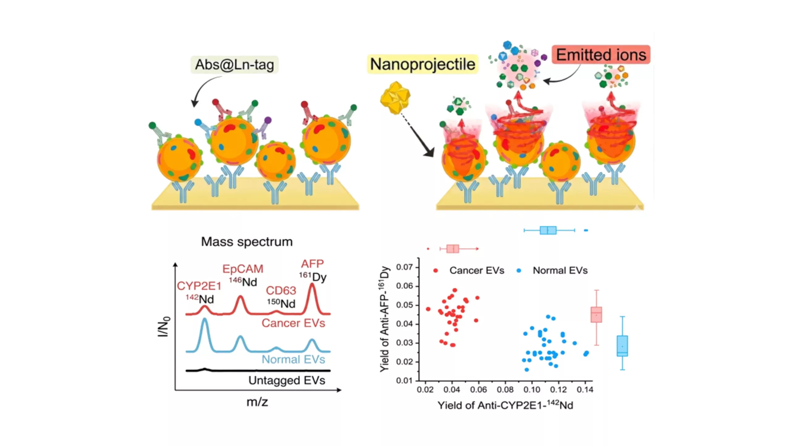
Nanoprojectile SIMS (NP-SIMS) has measured four protein biomarkers on individual extracellular vesicles from liver cells, telling those released by cancer cells apart from those released by healthy ones.
Every cell in the body sheds extracellular vesicles (EVs), tiny membrane-bound packages that carry proteins from the cell that made them. Because that cargo reflects where a vesicle came from, EVs have become a major focus of biomarker research, including in cancer.
Studying them is harder than it sounds. No two EVs are quite the same, yet most analytical methods measure millions of them at once and report an average. Whatever makes one vesicle different from its neighbour is lost in the result. Single extracellular vesicle analysis gets around this by looking at each vesicle on its own.
That was the aim of a team from Texas A&M University, Mayo Clinic and California State University, who turned to nanoprojectile secondary ion mass spectrometry (NP-SIMS) to do it.
Why extracellular vesicle heterogeneity matters
Cells release EVs into blood and other body fluids, and each vesicle carries proteins from the cell it came from. That makes EVs attractive for biomarker research and liquid biopsy studies.
The difficulty is that EVs vary a great deal. A single sample contains vesicles from different cell types, at different stages, carrying different combinations of proteins. Common methods such as western blotting and ELISA measure the whole sample at once. The signal from a small but important subpopulation can disappear into the average.
Single extracellular vesicle analysis addresses this by asking a different question: not how much of a protein is in the sample, but which vesicles carry it and what else they carry alongside it.
How the study worked
The researchers started with a simple comparison: EVs from normal human hepatocytes and EVs from HepG2 liver cancer cells. On size alone, you would struggle to tell them apart. Nanoparticle tracking analysis measured mean diameters of 112 ± 21 nm for the normal EVs and 119 ± 28 nm for the cancer EVs.
If size could not separate the two, the proteins on their surface might. The question was how to read those proteins one vesicle at a time.
Tagging the EV biomarkers
The first step was to hold the vesicles in place. The team captured them on gold surfaces coated with anti-CD63 antibodies, checking each stage of the surface preparation with surface plasmon resonance. SEM images confirmed that individual EVs were spread across the surface.
Next, they labelled four proteins with antibodies carrying different lanthanide isotopes. Each tag has its own mass, so a single measurement can pick out all four markers together. To check the tags were binding where they should, the team ran controls with an isotype control antibody and with surfaces carrying no EVs. Both gave signals close to background.
What is NP-SIMS?
With the vesicles tagged, NP-SIMS could get to work. The instrument fires single Au400 gold clusters at the surface, at about 1 keV per atom. Each impact releases ions from an area roughly 10 nm across, far smaller than a single EV, and is recorded as its own mass spectrum.
Around a million of these spectra were collected per sample. Recording them separately has a useful consequence: when two tags appear in the same spectrum, the proteins they mark were sitting close together on the vesicle surface.
What the researchers found
All four tags were detected on the same samples, and the two types of EV carried quite different amounts of each marker. Cancer EVs had more than twice as much AFP as normal EVs, and less than half as much CYP2E1.
Biomarker | Mass tag | Markers detected, normal EVs | Markers detected, cancer EVs |
|---|---|---|---|
CD63 (general EV marker) | 150Nd | 1.6 × 10⁵ | 1.5 × 10⁵ |
CYP2E1 (liver enzyme) | 142Nd | 6.5 × 10⁵ | 2.7 × 10⁵ |
EpCAM (epithelial marker) | 146Nd | 3.8 × 10⁵ | 5.5 × 10⁵ |
AFP (liver cancer marker) | 161Dy | 2.6 × 10⁵ | 6.7 × 10⁵ |
When the researchers combined AFP, CYP2E1 and EpCAM in a tSNE analysis, the cancer and normal EVs fell into two clearly separate groups.
The co-detection data added another layer. On cancer EVs, AFP and CD63 were found together about 11 times more often than on normal EVs, above the level expected if the markers were positioned at random. In a 1:1 mix of cancer and normal EVs, that figure dropped to about half. Nanoflow cytometry, run as an independent check, confirmed the higher AFP levels in the cancer EVs.
The study used EVs from cultured cells rather than patient samples. Within that setting, it shows that NP-SIMS can measure several protein biomarkers on individual EVs and tell cancer-derived vesicles from normal ones, even when the two are almost the same size.
Reference:
Lee, S., Verkhoturov, D. S., Eller, M. J., Verkhoturov, S. V., Shaw, M. A., Gwon, K., Kim, Y., Lucien, F., Malhi, H., Revzin, A., & Schweikert, E. A. (2023). Nanoprojectile secondary ion mass spectrometry enables multiplexed analysis of individual hepatic extracellular vesicles. ACS Nano, 17(23), 23584–23594. Read full paper here
Alba Scientific brings Bienne Technology’s Orion NP-SIMS to European researchers, backed by local application expertise, training, and technical support. We also provide sample measurement services so you can test performance on real samples before buying. Get in touch to discuss your research needs.
Follow us on linkedin