
Nanoprojectile secondary ion mass spectrometry:
NP- SIMS
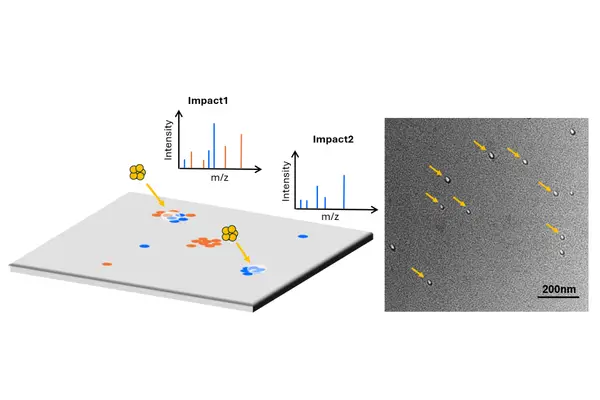
Nanoprojectile secondary ion mass spectrometry (NP-SIMS) is an event-by-event surface analysis technique that bombards a sample with individual, highly charged nano-projectiles, capturing a separate mass spectrum from every impact rather than averaging a beam over the surface.
Introduction
Conventional surface analysis methods, such as TOF-SIMS, use a continuous ion beam and produce an average chemical signal from the area analysed. While effective for bulk measurements, this can hide important differences between nearby nanoscale regions.
Nanoprojectile SIMS (NP-SIMS) takes a different approach by firing individual nanoparticles, usually gold clusters, onto the surface one at a time. Each impact creates a tiny crater and releases molecules that are measured as a separate mass spectrum linked to that exact location.
By collecting millions of individual measurements, NP-SIMS builds detailed chemical maps of surfaces at the nanoscale, revealing variations that would otherwise be lost in averaged measurements.
Nanoprojectile SIMS (NP-SIMS) replaces the continuous or pulsed ion beam used in conventional TOF-SIMS with a stream of individual, highly charged nanoprojectiles, typically gold clusters such as Au₄₀₀⁴⁺ or Au₂₈₀₀⁸⁺. Each projectile impact is separated in space and time and recorded as an individual event, allowing the resulting secondary ions to be linked directly to a specific nanoscale location.
Each impact delivers high energy to a very small surface area, generating a strong burst of secondary ions from a crater typically around 10–20 nm in diameter, while producing less fragmentation than conventional atomic ion bombardment. At impact rates of around 1 kHz, NP-SIMS can collect millions of individual mass spectra in a single analysis.
This event-by-event approach preserves nanoscale chemical variations that are lost in averaged measurements. The data can be analysed as a whole for statistical information or grouped by composition to reveal chemical heterogeneity across the surface. As each impact samples only a tiny fraction of the analysed area, the technique leaves most of the sample surface intact for complementary methods such as SEM or AFM.
Each nanoprojectile impact creates a localised, high-energy collision cascade. Gold clusters containing hundreds to thousands of atoms, accelerated to around 1 keV per atom, deposit their energy into a nanoscale surface volume, creating a highly excited transient region.
Unlike monatomic ion bombardment, cluster impacts concentrate energy near the surface, increasing secondary ion yield while limiting penetration depth. This produces a sampled volume that remains genuinely nanoscale, typically defined by craters around 10–20 nm in diameter.
By resolving each impact individually, NP-SIMS provides direct access to impact-to-impact variations in sputtering yield, ion emission and cluster–surface collision dynamics. Time-resolved measurements at kHz repetition rates also enable analysis of co-emitted species from single collision events, providing insight into energy transfer and fragmentation processes during cluster sputtering.
Nanoprojectile SIMS (NP-SIMS) provides detailed molecular surface information with lower fragmentation than conventional atomic ion SIMS. By distributing impact energy across a cluster of atoms, NP-SIMS improves the survival and detection of larger molecular and quasi-molecular ions, making it particularly useful for polymers, organic residues and biomolecular surfaces.
Each impact produces its own mass spectrum, capturing co-emitted ions from the same nanoscale region. This provides direct insight into chemical co-localisation — showing which species are associated at the nanoscale rather than simply detected somewhere within the analysed area. The technique enables detailed studies of nanoparticle surface chemistry, ligand coverage, particle interfaces and surface-bound molecular assemblies.
Nanoprojectile SIMS (NP-SIMS) enables molecular analysis of individual biological nanostructures, including extracellular vesicles, lipid assemblies and surface-labelled nanoparticles. Rather than measuring an average signal from a population, each particle or nanoscale region produces its own mass spectrum.
By using mass-distinguishable labels, such as lanthanide-tagged antibodies, NP-SIMS can identify which surface markers are present together on the same individual particle. This reveals biological differences between particle subpopulations that can be hidden in bulk measurements, enabling detailed studies of vesicle heterogeneity, disease-related changes and nanoscale biomolecular organisation.
Key features
Areas of use
Nanoprojectile SIMS (NP-SIMS) is used when researchers need detailed surface chemistry information at the nanoscale, beyond what conventional averaged measurements can reveal.
- Mapping trace contaminants on semiconductor and microelectronic surfaces
- Checking the chemistry of nanoscale features in advanced lithography processes
- Studying the surface chemistry of catalysts at the level of individual active sites
- Characterising nanocomposites and thin-film materials feature by feature
- Analysing individual biological nanoparticles, such as extracellular vesicles, for disease markers
By recording each impact separately, NP-SIMS reveals chemical differences across a surface that would otherwise be hidden by averaging.
- Semiconductor metrology : direct localisation and identification of nanoscale contaminants and process residues without averaging across device features.
- EUV lithography : resist film process control and defect analysis with nanometre-scale chemical resolution.
- Heterogeneous catalysis : surface chemical characterisation of catalytic materials at the scale of individual active sites.
- Materials science : chemical analysis of nanocomposites and multiphase materials, resolving individual domains rather than bulk composition.
- Multi-user facilities : a shared analytical tool for nanoscale chemical imaging across multiple research groups and applications
- Cluster–surface collision physics : direct, impact-resolved study of sputtering yield, energy deposition and fragmentation pathways in cluster-induced collision cascades
- Nanoscale metrology: sub-15 nm chemical resolution for studying interfaces, boundaries and adjacent nanostructures.
- Coincidence and co-emission analysis: identification of species emitted together from individual collision events, providing insight into energy partitioning and fragmentation mechanisms.
- Thin film and interface characterisation : nanoscale chemical profiling of lateral and buried interfaces, including capped and multilayer structures.
- Ligand shell characterisation : determining organic ligand composition, coverage and particle-to-particle variation on individual nanoparticles.
- Nanoparticle population analysis : distinguishing particles with similar core sizes but different surface chemistries.
- Particle–particle and particle–substrate interfaces : probing interfacial chemistry at sub-5 nm length scales beyond the reach of averaged surface analysis methods.
- Polymer and organic surface analysis : detecting larger, intact molecular ions from soft and organic materials through reduced fragmentation.
- Extracellular vesicle profiling : multiplexed surface marker analysis of individual EVs for biomarker discovery and disease research, including cancer diagnostics.
- Single-nanoparticle bioanalysis : resolving biological variation within nanoparticle populations that would appear as a single average in bulk measurements.
- Lipidomics and membrane chemistry : mapping lipid composition and membrane chemistry at the nanoscale on individual vesicles and membrane fragments.
- Label-free and tagged biomarker detection : combining native molecular analysis with antibody-based mass tags for targeted identification of biological markers.
Application areas
Deep reading
- Nanoprojectile secondary ion mass spectrometry for nanometrology of nanoparticles and their interfaces.
- Nanoprojectile secondary ion mass spectrometry for analysis of extracellular vesicles.
- Nanoprojectile secondary ion mass spectrometry enables multiplexed analysis of individual hepatic extracellular vesicles.